New Step in Personalized Drug Design: TTR Mutations Being Mapped
An international research team is uncovering new molecular mechanisms associated with pathogenic mutations in the transthyretin (TTR) protein, which cause transthyretin amyloidosis (ATTR). The results, obtained through a new methodological approach, pave the way for the development of drugs specifically designed for disease-related protein variants with higher therapeutic potential.
An international research team has identified new molecular mechanisms associated with pathogenic mutations in transthyretin (TTR) protein, which causes transthyretin amyloidosis (ATTR). Results obtained through a novel methodological approach open the way for developing drugs specifically designed for disease-associated protein variants with greater therapeutic potential.
The study was published in the Proceedings of the National Academy of Sciences. The research was led by researchers from the Institute of Biotechnology and Biomedical Research at Barcelona Autonomous University (IBB-UAB) and Washington University in St. Louis.
Understanding transthyretin and ATTR
Transthyretin (TTR) is a protein produced primarily in the liver and to a lesser extent in the brain. Certain genetic mutations cause TTR to misfold and aggregate, leading to the accumulation of TTR in the form of amyloid fibrils in various tissues. This accumulation leads to a group of progressive and fatal clinical disorders known as transthyretin amyloidosis (ATTR), which can affect the nervous system, heart, and other vital organs.
High-resolution X-ray diffraction studies have determined more than 300 TTR structures; however, these provide a static image of the protein and do not capture the effects of pathogenic mutations on TTR's stability and conformation. Small molecules (binding ligands) have been developed to counteract the effects of these mutations; however, currently approved drugs have generic activity and do not provide specific therapeutic response for different phenotypic variants of the disease. This situation demonstrates the need to design new stabilizers adapted to each specific mutation.
New research methods and findings
In this study, researchers focused on a new methodological approach to analyzing pathogenic TTR mutations. This approach enabled them to obtain important findings about the conformational changes caused by pathogenic TTR mutations and particularly how stabilizing ligands can counteract these effects. The method presents a dynamic mechanism of action that can be compared to a "film" rather than a static image.
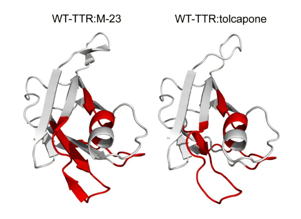
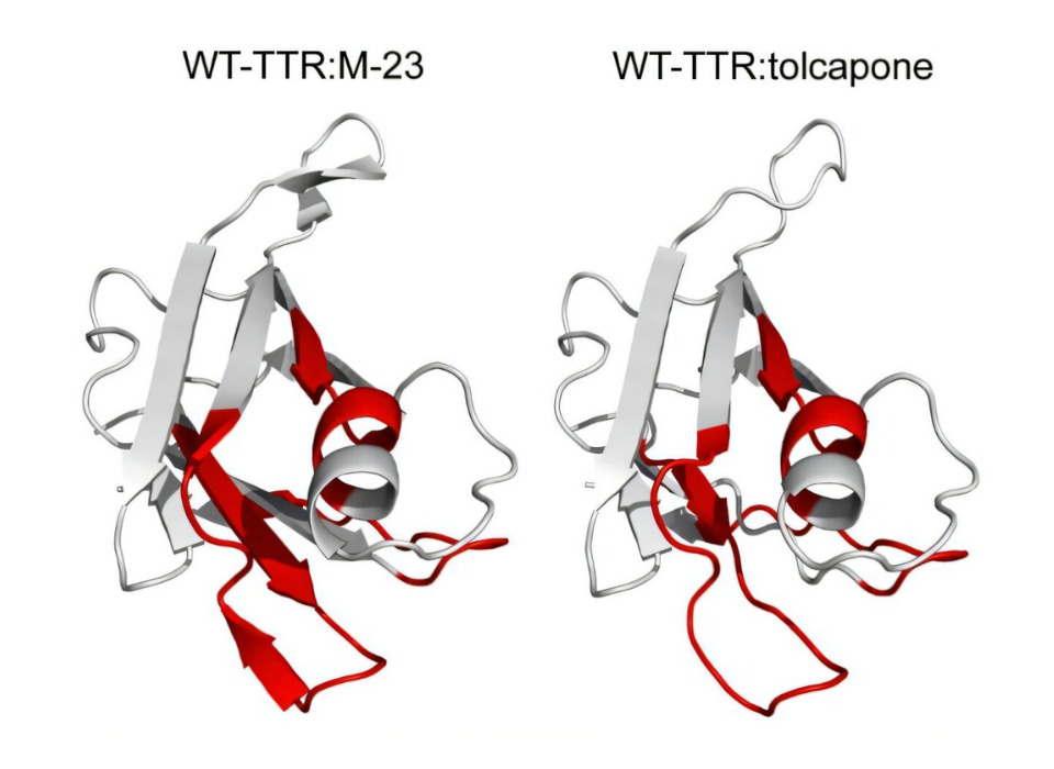
Irantzu Pallarès, researcher at the Protein Folding and Conformational Diseases Group at UAB, explains: "By combining mass spectrometry (MS), hydrogen-deuterium exchange (HDX), and fast photochemical oxidation of proteins (FPOP), we were able to observe conformational changes induced by both mutations and ligand binding that were not visible with X-ray crystallography."
Salvador Ventura, IBB-UAB researcher, professor at the Department of Biochemistry and Molecular Biology, and director of Parc Taulí Research and Innovation Institute (I3PT), states: "We uncovered previously hidden destabilization mechanisms, which creates new ground for designing specific stabilizers with significantly increased therapeutic potential for each mutation. Therefore, in the design of new ligands, the dynamic properties of each pathogenic variant of TTR should be taken into account."
The researchers conclude that the inclusion of MS-based techniques in ATTR drug discovery will accelerate the development of inhibitors that can prevent the aggregation of disease-associated variants with much greater precision.
Source - Francisca Pinheiro et al, Mass spectrometry footprinting reveals how kinetic stabilizers counteract transthyretin dynamics altered by pathogenic mutations, Proceedings of the National Academy of Sciences (2025). DOI: 10.1073/pnas.2519908122 / Journal information: Proceedings of the National Academy of Sciences / https://phys.org/news/2026-01-method-reveals-mutations-transthyretin-amyloidosis.html
Gallery